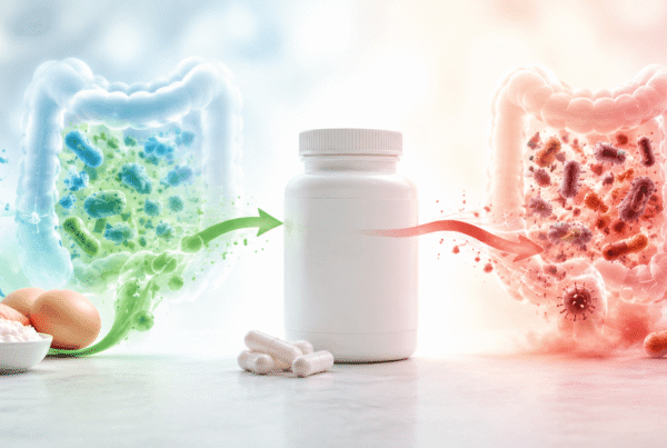

Yes. Inflammatory signalling molecules act directly on the hypothalamus and pituitary — the control centres sitting above your thyroid, adrenal and reproductive hormones. When inflammation is present, it changes the instructions being sent to all three. This means a hormone result can be genuinely abnormal while the hormone glands themselves are working perfectly well, and it’s one of the more common reasons hormone treatment doesn’t hold.
How can inflammation affect hormones at all?
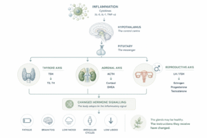
Hormone production isn’t controlled by the glands themselves. It runs top-down: the hypothalamus signals the pituitary, the pituitary signals the thyroid, adrenals and ovaries or testes, and those glands produce the hormones you measure on a blood test.
Immune signalling molecules — cytokines such as IL-6, IL-1 and TNF-α — act on that control system, not just on injured tissue. Receptors for IL-6 are present on pituitary cells and on adrenal cells directly¹, which is how an immune signal ends up changing an endocrine output.
The effect is not a malfunction. It’s a coordinated response: when the body is dealing with infection or injury, it down-regulates processes that can wait — reproduction in particular — and shifts resources toward the immune response. The problem arises when the inflammation doesn’t resolve, and a short-term adjustment becomes the ongoing state.
What does inflammation do to cortisol and the adrenals?
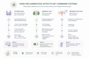
Inflammatory cytokines activate the HPA axis directly. In human studies, giving IL-6 produced marked and prolonged rises in ACTH and cortisol², and IL-6 turns out to be a particularly potent stimulator of the human adrenal axis³.
Two things follow from that.
First, cortisol output rises to meet the demand. Over time, sustained demand changes the pattern of what the adrenals produce — including DHEA, which is made in the same glands and often falls when the system has been under prolonged load.
Second, the axis adapts. In the same human research, the ACTH response to IL-6 was noticeably blunted after a week of repeated exposure². The system resets to a new equilibrium. This is why a single cortisol reading taken well into a chronic process can look unremarkable and still not reflect what’s actually happening — and why measuring both free and metabolised cortisol tells you more than either alone.
What does inflammation do to the thyroid?
This one is well described in the literature and has its own name: non-thyroidal illness syndrome, also called low T3 syndrome or sick euthyroid syndrome⁴.
The characteristic pattern is falling T3, rising reverse T3, and a TSH that stays normal or inappropriately unchanged⁵. In other words, the thyroid gland is fine and TSH looks acceptable, but the active hormone isn’t being produced.
The mechanism is in the conversion enzymes. Most circulating T3 doesn’t come from the thyroid — it’s made by converting T4 in peripheral tissue, using deiodinase enzymes. Inflammation changes how those enzymes behave: IL-6 has been shown to inhibit T3 production by the type 1 and type 2 deiodinases⁶, while type 3 deiodinase activity — which inactivates thyroid hormone and generates reverse T3 — increases⁵.
The practical consequence is that thyroid hormone can be low in tissue while a standard blood panel reads as normal. That’s a large part of why someone can have every hallmark of low thyroid function and be told their thyroid is fine.
One caveat: most of this research comes from critical illness and hospitalised patients, where the inflammatory signal is large. Whether low-grade chronic inflammation produces the same pattern to the same degree is less firmly established, though the mechanism is the same one.
What does inflammation do to reproductive hormones?
Reproduction is the first thing the body de-prioritises when conditions look unsafe, and inflammation is one of the signals it reads that way.
Cytokines act on GnRH neurons in the hypothalamus — the cells that set the pulse driving the entire reproductive axis⁷. Suppress that pulse and everything downstream follows: LH and FSH fall, and ovarian or testicular hormone production falls with them. This is the mechanism behind functional hypothalamic amenorrhoea, where periods stop or never start despite normal reproductive anatomy.
The effect isn’t only central. In men, gut-derived bacterial endotoxin has been shown to reduce testosterone production directly at the Leydig cell⁸, by disrupting the mitochondria those cells need for steroid synthesis⁹.
Where is the inflammation coming from?
Often the gut, and this is the part that gets missed.
The intestinal lining separates a large bacterial population from the bloodstream. When that barrier becomes more permeable, bacterial endotoxin — lipopolysaccharide, or LPS — passes into circulation and triggers a systemic inflammatory response. This has been proposed as a key inflammatory trigger in male hypogonadism¹⁰, and in a controlled human study, endotoxin exposure correlated with both raised IL-6 and lower testosterone⁸.
So a hormone problem can begin as a gut problem. Chronic infection, bacterial or fungal overgrowth, and intestinal permeability all keep inflammatory signalling elevated, and that signal reaches the hypothalamus and pituitary regardless of where it started.
Why do hormone tests look normal when this is happening?
Three reasons, and they compound.
The glands aren’t the problem. Testing usually measures gland output. If the issue is upstream signalling, the gland can be responding appropriately to a poor instruction, and every number can sit inside its range.
Reference ranges are wide. They describe a population, not a person. A result can be technically normal and still be wrong for someone’s age or circumstances — DHEA-S in a teenager being the clearest example.
The relationship between markers carries the information. Low T3 with high reverse T3 and a normal TSH is a recognisable pattern. Read one marker at a time and the pattern disappears.
What can be done about it?
The logic is straightforward even when the case isn’t: find the inflammatory driver, because treating the hormone downstream of an unresolved driver rarely holds.
- Look for the source. Gut infection or overgrowth, intestinal permeability, chronic infection, autoimmune activity, and mould or biotoxin exposure are the common candidates.
- Measure inflammation, not just hormones. Inflammatory markers alongside a hormone panel change the interpretation of both.
- Test the axis, not the gland. A full thyroid panel including T3 and reverse T3 tells you more than TSH. Cortisol measured across the day with metabolites tells you more than a single reading.
- Expect hormone results to move once the driver is addressed. If they don’t, that’s useful information too — it suggests something else is going on.
This isn’t a reason to ignore hormone treatment where it’s needed. It’s a reason to know whether you’re treating the problem or its shadow.
Frequently asked questions
Can inflammation cause low thyroid symptoms with normal test results? Yes. Inflammatory cytokines alter the enzymes that convert T4 into active T3, lowering T3 and raising reverse T3 while TSH stays normal. This pattern is well described as non-thyroidal illness syndrome.
Can gut problems cause hormone problems? Indirectly, yes. Increased intestinal permeability allows bacterial endotoxin into circulation, which triggers systemic inflammation. That inflammatory signal acts on the hypothalamus and pituitary, which control thyroid, adrenal and reproductive hormone output.
Does inflammation affect cortisol? Yes. Inflammatory cytokines including IL-6 stimulate the adrenal axis directly and raise cortisol. With prolonged exposure the axis adapts and settles at a new equilibrium, which is why a single cortisol reading can be misleading in a chronic process.
Can inflammation stop your periods? It can contribute. Inflammatory signalling suppresses the GnRH neurons that drive the reproductive axis, reducing the downstream hormone production needed for a cycle. This is one mechanism behind functional hypothalamic amenorrhoea.
Should I test for inflammation or potential causes of inflammation as well as hormones? Testing both together is more informative than either alone, because inflammation changes how a hormone result should be read.
References
- Turnbull, A. V. & Rivier, C. L. (1999). Regulation of the hypothalamic-pituitary-adrenal axis by cytokines: actions and mechanisms of action. Physiological Reviews, 79(1), 1–71. doi:10.1152/physrev.1999.79.1.1
- Mastorakos, G., Chrousos, G. P. & Weber, J. S. (1993). Recombinant interleukin-6 activates the hypothalamic-pituitary-adrenal axis in humans. Journal of Clinical Endocrinology & Metabolism, 77(6), 1690–1694. PMID: 8263159
- Bethin, K. E., Vogt, S. K. & Muglia, L. J. (2000). Interleukin-6 is an essential, corticotropin-releasing hormone-independent stimulator of the adrenal axis during immune system activation. PNAS, 97(16), 9317–9322. doi:10.1073/pnas.97.16.9317
- An update on non-thyroidal illness syndrome. Journal of Endocrinological Investigation (2021). PMC8285315
- The molecular basis of the non-thyroidal illness syndrome. Journal of Endocrinology (2015), 225(3), R67–R81.
- New insights toward the acute non-thyroidal illness syndrome. Frontiers in Endocrinology (2012). PMC3356062
- Effect of inflammation on female gonadotropin-releasing hormone (GnRH) neurons: mechanisms and consequences. International Journal of Molecular Sciences (2020), 21(2), 529. doi:10.3390/ijms21020529
- Tremellen, K. et al. (2018). Endotoxin-initiated inflammation reduces testosterone production in men of reproductive age. American Journal of Physiology – Endocrinology and Metabolism. doi:10.1152/ajpendo.00279.2017
- Allen, J. A. et al. (2005). Bacterial endotoxin lipopolysaccharide and reactive oxygen species inhibit Leydig cell steroidogenesis via perturbation of mitochondria. Endocrine, 25(3), 265–275. PMID: 15758255
- Tremellen, K. (2016). Gut Endotoxin Leading to a Decline IN Gonadal function (GELDING) — a novel theory for the development of late onset hypogonadism in obese men. Basic and Clinical Andrology, 26, 7. doi:10.1186/s12610-016-0034-7